Comparative histopathological studies of selected organs of Oreochromis niloticus (Linnaeus, 1758) from Igun and Opa Reservoirs, Southwestern Nigeria
DOI:
https://doi.org/10.55779/nsb14211235Keywords:
fish organs, histopathology, Igun and Opa ReservoirsAbstract
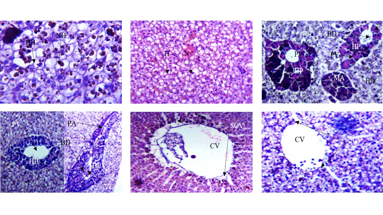
Heavy metals have been reported to have negative impacts on the histology of fishes. In this study, the impact of heavy metal bioaccumulation on the gills, muscle and liver of Oreochromis niloticus from Opa and Igun reservoirs was determined by histological methods. This was with a view to checking for possible alterations on fish organs. Live fish samples were collected from Opa and Igun reservoirs and identified in the laboratory. Histological analyses were carried out on the organs and their photomicrographs taken using digital binocular compound LED microscope. The gills of O. niloticus from Opa and Igun reservoirs showed hyperplasia of secondary lamellae and hypertrophy of primary lamellae while shortening and edema of secondary lamellae were observed in O. niloticus from Opa reservoir only. Muscular atrophy and degeneration were revealed in the muscle of fish from the two reservoirs with muscular splitting in O. niloticus from Igun reservoir. The liver of O. niloticus from Opa reservoir showed vascular congestion in the bile duct compared to O. niloticus from Igun reservoir which showed hepatopancreas degeneration, melanomacrophages aggregates, nucleus and hepatocytes hypertrophy. In conclusion, histopathological alterations were more severe in the organs of O. niloticus from Igun reservoir compared to that from Opa reservoir.
Metrics
References
Adesulu EA, Sydenham DHJ (2007). The freshwater fishes and fisheries of Nigeria. Macmillan Nigeria Publishers Limited., Ibadan, pp 397.
Agius C, Roberts RJ (2003). Melanomacrophage centers and their role in fish pathology. Journal of Fish Diseases 26:499-509. https://doi.org/10.1046/j.1365-2761.2003.00485.x
Arawomo GAO (1987). The fish fauna of the rivers in the New Federal Capital Territory, Abuja, Nigeria. Ife Journal of Science 2:37-43.
Au DWT (2004). The application of histo-cytopathological biomarkers in marine pollution monitoring: a review. Marine Pollution Bulletin 48:817-834. https://doi.org/10.1016/j.marpolbul.2004.02.032
Bancroft JD, Cook HC (1994). Manual of histological techniques and their diagnostic application. Churchill Livingstone, London, pp 289-305. https://doi.org/10.1002/path.1711450410
Bernet D, Schmidt H, Meir W, Burkhardt-Holm P, Wahli T (1999). Histopathology in fish: proposal for a protocol to assess aquatic pollution. Journal of Fish Diseases 22:25-34. https://doi.org/10.1046/j.1365-2761.1999.00134.x
Bhuvaneshwari R, Padmanaban K, Babu Rajendran R (2015). Histopathological alterations in muscle, liver and gill, tissues of zebra fish Danio rerio due to environmentally-relevant concentrations of organochlorine pesticides (OCPs) and heavy metals. International Journal of Environmental Research 9(4):1365-1372. https://doi.org/10.22059/IJER.2015.1029
Birungi Z, Masola B, Zaranyika MF, Naigaga I, Marshall B (2007). Active biomonitoring of trace heavy metals using fish (Oreochromis niloticus) as bioindicator species. The case of Nakivubo wetland along Lake Victoria. Physics and Chemistry of the Earth 32(15-18):1350-1358. http://dx.doi.org/10.1016/j.pce.2007.07.034
Camargo MMP, Martinez CBR (2007). Histopathology of gills, kidney and liver of a Neotropical fish caged in an urban stream. Neotropical Ichthyology 5(3):327-336. https://doi.org/10.1590/S1679-62252007000300013
Eller LL (1975). Gill lesions in freshwater teleosts. In: Ribelin WE, Migaki G (Eds). The Pathology of Fishes. University of Wisconsin Press, Madison, pp 305-330.
Evans DH (1993). Osmotic and ionic regulation. In: Evans DH (Ed). The Physiology of Fishes. Boca Raton, FL: CRC, pp 315-341.
Farkas A, Salanki J, Specziar A (2002). Relation between growth and the heavy metal concentration in organs of bream Abramis brama L. populating Lake Balaton. Archive Environmental Contamination Toxicology 43:236-243. https://doi.org/10.1007/s00244-002-1123-5
Fuggle RF, Rabie MA (1994). Environmental Management in South Africa. Cape Town, Juta, pp 1142. https://doi.org/10.1080/0035919X.2011.593204
Gaber HS (2013). Fish health as a biomarker for the condition of Lake Nasser. Journal of Applied Sciences Research 9(11):5794-5810.
Gernhofer M, Pawet M, Schramm M, Muller E, Triebskorn R (2001). Ultra-structural biomarkers as tools to characterize the health status of fish in contaminated streams. Journal of Aquatic Ecosystem, Stress and Recovery 8:241-260. https://doi.org/10.1023/A:1012958804442
Hadi AA, Alwan SF (2012). Histopathological changes in gills, liver and kidney of fresh water fish, Tilapia zillii, exposed to aluminum. International Journal of Pharmacy and Life Sciences 3(11):2071-2081.
Lawal OA, Komolafe OO (2012): Concentrations of heavy metals in three Tilapine species of an abandoned Gold Mine Reservoir in Igun, Nigeria. Nigerian Journal of Fisheries 9(2):581-585.
Malik GM, Raval HV, Ahmed Khali HK (2012). Toxic effects of effluent on mortality and behaviour changes on fresh water fish Poecilia reticulate. Journal of Environmental Research and Development 7(2A):1036-1039.
Marchand MJ, Van Dyk JC, Pieterse GM, Barnhoorn IE, Bornman MS (2009). Histopathological alterations in the liver of the sharp tooth catfish, Clarias gariepinus from polluted aquatic systems in South Africa. Environmental Toxicology 24:133-147. https://doi.org/10.1002/tox.20397
Mazon AF, Monteiro EAS, Pinheiro GHD, Fernandes MN (2002). Hematological and physiological changes induced by short-term exposure to copper in the freshwater fish, Prochilodus scrofa. Brazilian Journal of Biology 62(4A):621-631. https://doi.org/10.1590/s1519-69842002000400010
Mohamed FAS (2009). Histopathological Studies on Tilapia zillii and Solea vulgaris from Lake Qarun, Egypt. World Journal of Fish and Marine Sciences 1(1):29-39.
Ochieng GM, Seanego ES, Nkwonta OI (2010). Impacts of mining on water resources in South Africa: A review. Scientific Research and Essays 5(22):3351-3357. https://doi.org/10.5897/SRE.9000572
Olabanji IO, Oluyemi EA (2014). Preliminary assessment of heavy metal pollution of Opa Reservoir, Ile- Ife, Southwest Nigeria using Mormyrus rume and Tilapia zillii. Ife Journal of Science 16(1):35-43.
Osman AGM, Al-Awadhi RM, Harabawy AS, Mahmoud UM (2010). Evaluation of the use of protein electrophoresis of the African catfish Clarias gariepinus (Burchell, 1822) for biomonitoring. Aquatic Pollution and Environmental Research Journal 4:235-243. https://doi.org/10.3923/erj.2010.235.243
Reed W, Burchad T, Hopson AJ, Jenness J, Yaro I (1967). Fish and fisheries of Northern Nigeria. Ministry of Agriculture, Northern Nigeria, pp 226.
Reddy PB, Baghel BS (2012). Impact of industrial waste water on the Chambal River and biomarker responses in fish due to pollution at Nagda, M.P. India. DAV International Journal of Science 1(1):86-91. https://doi.org/10.13140/RG.2.1.2701.6722
Reddy PB, Rawat SS (2013). Assessment of aquatic pollution using histopathology in fish as a protocol. International Research Journal of Environment Sciences 2(8):79-82.
Santos TCA, Gomes V, José M, Passos ACR, Rocha AJS, Salaroli RB, Van Ngan P (2011). Histopathological alterations in gills of juvenile Florida pompano Trachinotus carolinus (Perciformes, Carangidae) following sublethal acute and chronic exposure to naphthalene. Pan-American Journal of Aquatic Sciences 6(2):109-120.
Simonato JD, Guedes CLB, Martinez CBR (2008). Biochemical, physiological, and histological changes in the neotropical fish Prochilodus lineatus exposed to diesel oil. Ecotoxicology and Environmental Safety 69(1):112-120. https://doi.org/10.1016/j.ecoenv.2007.01.012
Stebbing ARD (1985). A possible synthesis. In Bayne BL (Ed). The Effects of Stress and Pollution on Marine Animals. New York, Praeger.
Trewavas E (1983). Tilapiine fishes of the genera Sarotherodon, Oreochromis and Danakilia. British Museum of Natural History, London.
Wood CM (2001). Toxic responses of the gill. In: Schlenk D, Benson WH (Eds). Target Organ Toxicity in Marine and Freshwater Teleosts Organ. Taylor and Francis, London. Vol. 1. pp 1-89. https://doi.org/10.1201/9781315109244
Yogita D, Mishra A (2013). Histopathological alterations in gill and liver anatomy of freshwater, air breathing fish Channa Punctatus after pesticide Hilban (Chlorpyrifos) treatment. Advanced Bioresearch 4(2):57-62.
Downloads
Published
How to Cite
Issue
Section
License
Papers published in Notulae Scientia Biologicae are Open-Access, distributed under the terms and conditions of the Creative Commons Attribution License.
© Articles by the authors; licensee SMTCT, Cluj-Napoca, Romania. The journal allows the author(s) to hold the copyright/to retain publishing rights without restriction.
License:

![]()
Open Access Journal - the journal offers free, immediate, and unrestricted access to peer-reviewed research and scholarly work, due SMTCT supports to increase the visibility, accessibility and reputation of the researchers, regardless of geography and their budgets. Users are allowed to read, download, copy, distribute, print, search, or link to the full texts of the articles, or use them for any other lawful purpose, without asking prior permission from the publisher or the author.
![]()


.png)